There is no longer a need to do an automatic focus point by point, making it faster than ever!
ViewSharpTM provides a clear view of all of the sample’s surfaces in the field of view. The topography is extracted and then used during the Raman acquisition. The Raman signal measurements are performed point by point with Raman spectra acquisition for every point. This complete signal acquisition enables multivariable analyses, or any other kind of analysis, to extract useful information about the studied sample.
- Get an instant topography of the sample
- Acquire highest quality in Focus Raman images
- Just turn it on, and surf the surface
ViewSharpTM Examples:
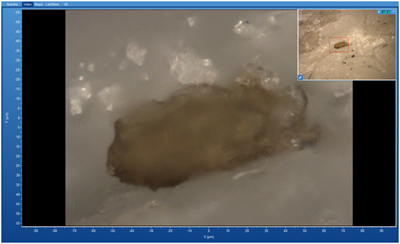
With ViewSharpTM (dolomite sample, 50x)
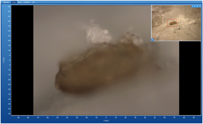
Without ViewSharpTM (dolomite sample, 50x)
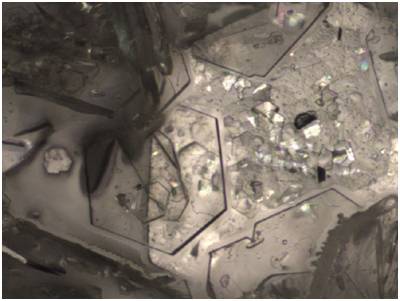
10x view of cystine sample with ViewSharpTM
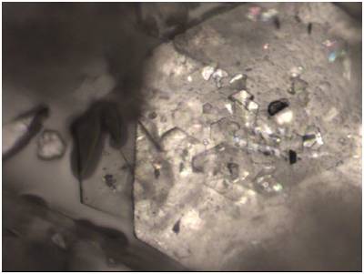
10x view of cystine sample without ViewSharpTM
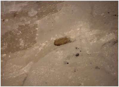
10x view of dolomite sample with ViewSharpTM
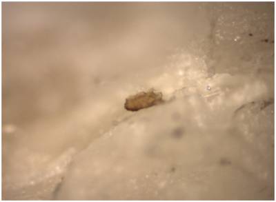
10x view of dolomite sample without ViewSharpTM
With ViewSharpTM , the topography of all sample points can be extracted:
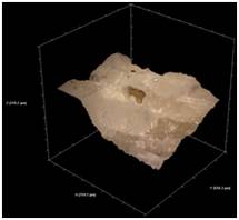 | 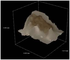 |
3D view of dolomite mineral sample at 10x | 3D view of dolomite mineral sample at 50x |
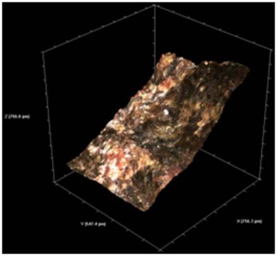
3D view of a rutile mineral sample, with a rough surface height of 766 µm, at 10x.
With ViewSharpTM, map the Raman image on top of the topography and get the highest quality Raman images.
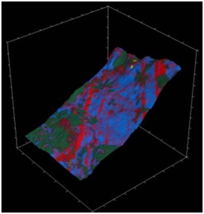
3D Raman mapping of a rutile mineral sample, with a rough surface height of 766 µm, at 10x. The rutile sample consists of two forms of TiO2 displayed in red and blue, anatase in yellow and organic contamination in green.
ViewSharpTM application is included is the groundbreaking EasyNavTM package.
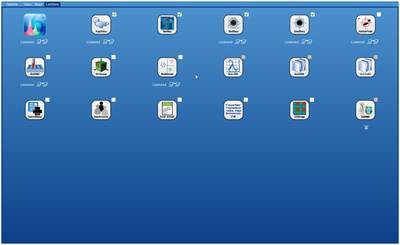
ViewSharpTM app is available in the LabStore of LabSpec 6 spectroscopy software suite:
