
HORIBA Scientific is fully aware of the power of spectroscopic imaging techniques, which provide valuable information to scientists seeking to understand interactions of matter and develop new materials. However the aesthetic qualities of the results should not be ignored either – in this Image Gallery we present some of the striking Raman images obtained on HORIBA Scientific Raman microscope systems, which can be appreciated by scientists and non-scientists alike.
If you would like to have your images included in the HORIBA Scientific Raman Image Gallery, please contact your local HORIBA Scientific office or representative, with a copy of the image and a brief summary of the work.
Geology | |||
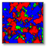 Mineral Section | 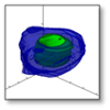 Fluid Inclusion | 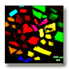 Carbonates | 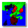 Granite |
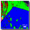 Rock Section | 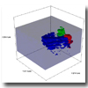 Fluid Inclusion 2 | ||
Pharmaceutics | |||
Life science | |||
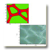 Wood | |||
Materials | |||
Expanded beads | Nano-indented Silicon | ||
Carbon | |||
Do you have any questions or requests? Use this form to contact our specialists.